April 20, 2023
Words & Photos by Jens Odegaard 

Mulan is meowling on the exam table. She’s just had an external splint on her sternum removed and been given the green light for final discharge from the Oregon State University Carlson College of Veterinary Medicine’s Lois Bates Acheson Veterinary Teaching Hospital. Dr. Brea Sandness, Dr. Bianca Reyes and fourth-year veterinary student Sean Calalang are delighted as they use moistened gauze to do a final cleaning of Mulan’s downy white chest fur. So is Dr. Ingrid Balsa who stands nearby.

Mulan is vivacious and ready for conquest in this great, big beautiful world. Six weeks ago, she was an undersized, weak, little, gray-and-white speck of a kitten carrying a huge weight on her chest.

Mulan was born with pectus excavatum. It’s a condition where the sternum and connecting tissues to the ribs are malformed, resulting in an irregularly flat or sunken chest. It’s a relatively common condition in kittens and puppies, and for many of them is mild enough that they are able to live healthy and unaffected lives. 

However, for more rare cases like Mulan, the deformity is severe enough that pressure is put on the heart and lungs, causing respiratory issues, exercise intolerance and a “failure to thrive,” Balsa says.

The key in these more severe – even life-threatening – cases, is to catch it early.

Going Rogue
Mulan is a rescue that was brought with her littermates to the Rogue Valley Humane Society in Grants Pass, Oregon when they were roughly seven weeks old. “She and her siblings seemed like healthy, normal kittens, so upon intake we scheduled her to get spayed. While she was under, as we were shaving her, we noticed there was a pretty pronounced divot in her sternum,” says Jaclyn Morris, the humane society’s office manager. “So we sent her off to get X-rayed. And that showed that she had a pretty severe bend to her rib cage, where it was almost like in danger of impaling or touching her heart and impacting her lungs so she wasn't able to take full complete deep breaths.”

In fact, the impact was so severe that Mulan even collapsed once after playing. In consultation with their local vet, the humane society made the decision to bring Mulan to Oregon State to access the expertise of veterinary specialists like Balsa and Sandness. “If it was left unchecked, she maybe wouldn’t have survived,” Morris says. “I am really grateful and lucky to work at a facility that is willing to kind of throw the kitchen sink at unique animals that come our way that need specific care. A lot of shelters and facilities may not have the luxury of putting all these resources into a cat like her, but we make that our philosophy — those are the animals that need us the most.” 

Procedural Approach
Dr. Brea Sandness, a small animal surgery faculty member and 2015 college alumna, took the lead on Mulan’s case with Dr. Bianca Reyes, a small animal surgical resident, assisting. Because cases like Mulan’s are rarely corrected (due to lack of resources or because they are caught too late), neither had performed this type of procedure. While working on Mulan, they’d also be teaching a team of veterinary students including fourth-year students Astrid Reyes and Selah Green and second-year student Marissa Patton. That’s the nature of a veterinary teaching hospital. Every patient visit and procedure is an educational experience. 

In preparing for the surgery, Sandness and Reyes focused on building the right team and knowledge base. 

Sandness reached out to Balsa to lend her expertise. Balsa is a board-certified small animal surgeon and a fellow in minimally invasive surgery (small animal soft tissue). Prior to joining Oregon State last fall, she was on the faculty at the UC Davis School of Veterinary Medicine. During her time there, she’d performed a handful of procedures for cases like Mulan’s. “To be able to work with these types of organizations that want to provide specialty care, it provides a great learning opportunity for some of these rarer cases for our residents to see and to learn from, so that they can then go on and hopefully help other animals,” Balsa says. “I’ve done a lot of work with shelters over my career and I've always found it a very gratifying experience with regards to their investment and care for the animals and then the learning that we get out of that.”  

Sandness then moved toward building out the rest of the game plan. “We only scheduled this when I knew that we would have the team, from anesthesia to the nurses to the surgeon scrubbing in with me, to make sure that we were going to be able to give this the best opportunity,” Sandness says. “Once that was confirmed … it was then researching the disease, pulling all the literature, seeing where did they run into complications, did they have any new techniques that were better than what I had read from before?”

There are two options to correcting pectus excavatum: invasive surgery to reconstruct the sternum or a minimally invasive approach where an external splint is sutured to the sternum. 

As a team, Sandness, Reyes and Balsa opted for the minimally invasive approach. “We were lucky in that she was young and still had a lot of flexibility in her bones as they were mineralizing,” Balsa says. “So we were able to do the less invasive external splint coaptation as opposed to some of the more invasive, either internal bracing or removal of some of the bones. Once we lose that flexibility and bones mineralize in place, we have to go to those more invasive things.” 

Eva Hayden, certified veterinary technician, shaves Mulan's chest in preparation for the procedure while fourth-year veterinary student Selah Green looks on. 

Drs. Sandness, Reyes and Balsa determining the rough outline and size of the splint prior to the procedure. 

Drs. Reyes, Sandness and Balsa outline the procedure before getting started. 


Saving Sutures 
With Mulan’s chest shaved and prepped for surgery, Sandness, Reyes and Balsa got to work. Step one was molding the plastic splint that would sit on the outside of Mulan’s chest. They cut the basic shape, then heated the plastic with hot water. Now malleable, the team formed the splint to the ideal contour for Mulan’s sternum to conform to over the next six weeks.

After a few minutes to cool, the splint was set and ready for attachment. 

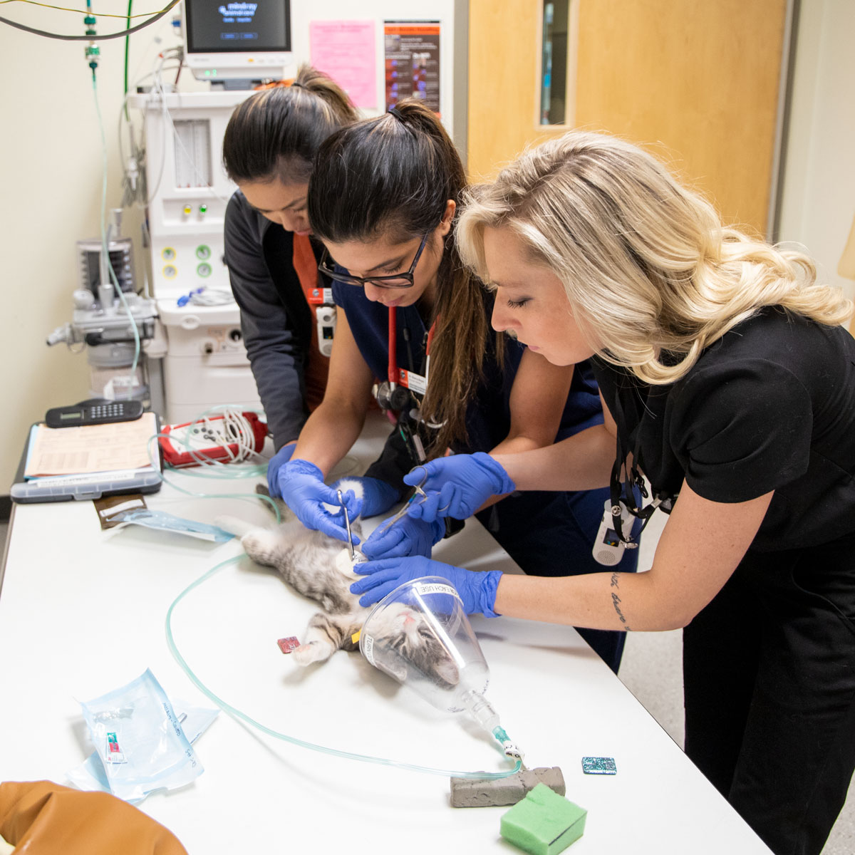
Next, was the critical part. Sandness steadied her hands, gripped a curved suturing needle with forceps and pierced Mulan’s skin. “Three millimeters was the margin of error between the surface of the skin to the inside of the sternum to get the suture around,” Sandness says. Take two credit cards, and lay them flat on top of each other. That’s about three millimeters thick. If Sandness went too deep, she risked piercing Mulan’s heart or lungs. 

She slid the needle down and around the sternum, drawing the suture through the layers of the skin, under the bones and then back up and out the other side. The suture was then cut, leaving several inches hanging out of each side, ready to be laced to the splint. Once, twice … six times, Sandness repeated the process, drawing the suture through as Balsa offered words of guidance and modeled the technique. 

Reyes observed and then grasped the needle for herself to do two sutures as Sandness offered guidance. “I was extremely happy, and I am very grateful to be part of this procedure and the program itself, both under the mentorship of Sandness and Balsa,” Reyes says. “This is a procedure that I've never seen before. I've read and heard about it, but I was very excited to finally be able to firsthand treat and help one of these congenital conditions.” 

Dr. Sandness listens as Dr. Balsa offers advice at the beginning of the procedure. 

Dr. Sandness starts the first suture. 

Dr. Balsa (center) demonstrates technique to Dr. Reyes (left) and Dr. Sandness. 

Dr. Sandness threads the suture through the splint. 

Dr. Reyes pulls the suture ends tight while Dr. Sandness holds the splint. 

Dr. Sandness and Dr. Reyes work together in threading the sutures through the splint. 


Eight suture laces now ran down beneath the sternum and back out through the skin on each side, ready to be cinched to the splint. 

With a straight needle, Sandness then punched eight holes down each side of the narrow splint.

To thread the suture through the holes, Balsa offered a pro-tip: insert a straight hollow needle, the kind you’d use to administer a shot, through the hole, then simply insert the end of the suture through the hollow needle and push it through. Genius. Experience.

With each suture end now threaded through a matching hole in the splint, it was simply a matter of cinching them up tightly. This drew the splint down securely against the outside of Mulan’s skin and the sternum up tightly against the internal side.

The end result looked like an external breastplate sewn tightly to Mulan’s chest.

Successful Six 
For the next six weeks, Mulan healed, returning to the teaching hospital for three checkups to make sure everything looked healthy and stayed clean. Then it was one last trip for the final exam and X-rays to ensure everything looked good to go. 

With thumbs-up from the diagnostic imaging team, Sandness, Balsa and Calalang snip the sutures free and pull the splint away from a much-changed Mulan. 

Thanks to the corrected growth of her sternum, Mulan’s heart and lungs are working uninhibitedly and Mulan is a big, healthy kitten. “Mulan is a pretty feisty kitten despite her start. She hasn't let that slow her down at all. She’s a mighty hunter, so she loves mice toys, she loves chasing little wand toys under the blanket. She also loves to play peekaboo. She's always watching, always paying attention, very playful, very energetic. And also a sweetheart when she's tired; she has her moments where she's really cute,” Morris says as she puts Mulan into her travel carrier after discharge. “But she is kind of a little Tasmanian devil, but I wouldn't have her any other way. She's really an awesome cat.” 
 

Dr. Reyes and Dr. Sandness remove the sutures from the splint while veterinary student Sean Calalang observes. 

Dr. Reyes removes the sutures while Dr. Sandness holds Mulan.

Drs. Balsa, Reyes and Sandness say their happy goodbyes to Mulan. 

Dr. Sandness holds Mulan while Dr. Reyes gives her an ear scratch. 

Dr. Balsa, Dr. Sandness, Dr. Reyes and Sean Calalang pose in the small animal clinic for a goodbye photo with Mulan.

Dr. Sandness holds Mulan on the exam table just before discharging her.